
Fig. 4. Assessment of the three tiers of oxidative stress in RAW 274.7 cells.
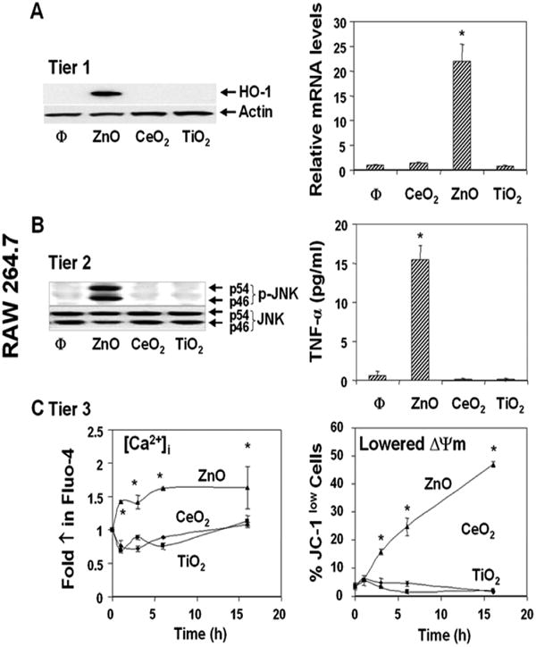
Cells were exposed to 25 μg/ml metal oxide NP for 1-16 hrs. The methodology for assessment of the hierarchical oxidative stress responses is described in Materials and Methods. (A) Induction of HO-1 expression (Tier 1) by immunoblotting and realtime PCR. (B) Jun kinase activation and TNF-α production in response to NPs (Tier 2). (C) Assessment of [Ca2+]i and mitochondrial membrane potential (ΔΨm) (Tier 3) using Fluo-4 and JC-1 fluorescent dyes, respectively. Flow cytometry was performed 1 - 16 hrs after the addition of the particles. The increase in [Ca2+]i can lead to cellular toxicity by triggering mitochondrial PT pore opening if the stories capacity of this organelle is exceeded. *p<0.01 compared with control. Data are representative of 3 separate experiments.