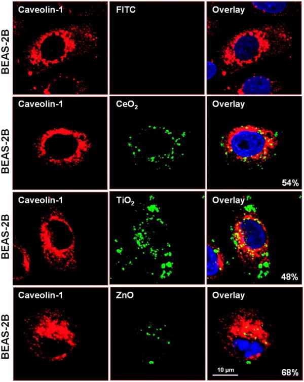
Fig. 7. Confocal microscopy to study the subcellular localization of FITC-labeled metal oxide NP in BEAS-2B cells.

Cells were exposed to 25 μg/ml FITC-labeled particles for 6 hr. After fixation and permeabilization, cells were stained with 1 μg/ml anti-caveolin-1 (BD BioSciences, San Jose, CA) and visualized using a Confocal 1P/FCS Inverted microscope. After merging of the red and green images, the % of cells with particles co-localizing with caveolae (composite yellow fluorescence) was quantified by Image J software.
