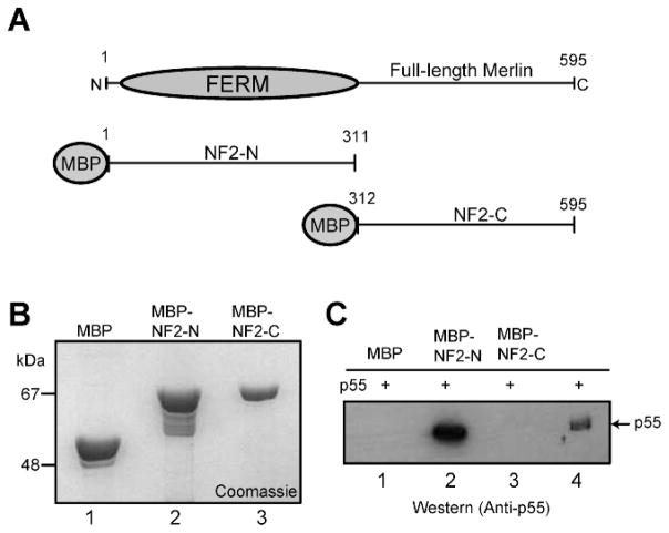
Figure 1.
In vitro binding between p55 and merlin. (A) Schematic representation of NF2 protein (merlin) constructs used for the binding assays. (B) Coomassie blue stained SDS-PAGE showing purified recombinant proteins. MBP, lane 1; MBP-NF2-N, lane 2; and MBP-NF2-C, lane 3. (C) Western blot based detection of p55 recovered by the MBP-beads pull-down assay. Purified recombinant His-p55 expressed in bacteria was incubated with MBP-fusions of NF2 protein immobilized on the beads. Lane 4 represents the input His-p55 protein used as a positive control.

