Figure 3.
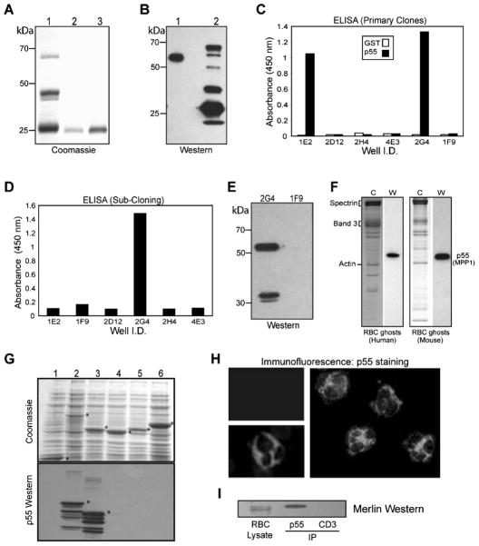
Generation of 2G4-monoclonal antibody against erythrocyte p55. (A) Coomassie staining of the GST-SH3-GUK domain of p55. Protein was expressed in bacteria and purified by glutathione-Sepharose beads. Lane 1: GST-SH3-GUK domain. Lanes 2 and 3: Elution of SH3-GUK polypeptide after thrombin cleavage. Polypeptides shown in lanes 2 and 3 were combined and injected into mice for immunization. (B) Western blot using serum from the immunized mice. Immune serum recognized one band in the human erythrocyte ghosts (lane 1) and multiple breakdown products in the GST-SH3-GUK domain construct (lane 2). (C) ELISA using GST and His-p55 proteins as immobilized antigens. Hybridoma supernatant from the primary screen identified two positive clones, 1E2 and 2G4. (D) ELISA screening of the subclones identified 2G4 as the only viable clone. (E) Western blotting shows that serum from the positive 2G4 clone recognizes p55 and its breakdown product in stored human erythrocyte ghosts. Supernatant from the negative clone 1F9 was included as a control. (F) Western blotting using 2G4 monoclonal identified a single 55 kDa band in both human and mouse erythrocyte ghosts. Coomassie staining is shown in the left panels. (G) Coomassie staining of GUK domain-constructs and the corresponding Western blots using 2G4 monoclonal antibody. Lanes 1, GST; 2, GST-SH3-GUK of p55; 3, GST-GUK of p55; 4, GST-GUK of human Dlg; 5, GST-GUK of human CASK; 6, GST-GUK of human ZO-1. The 2G4 monoclonal specifically recognizes the GUK domain of p55. (H) Immunofluorescence analysis of human peripheral blood cells. Intense p55 signal from neutrophils (two separate fields) was evident by staining with the 2G4 monoclonal antibody. The p55 staining in neutrophils was so strong that it dampened the signal from its uniform staining in the erythrocytes. The upper left panel indicates a negative sample where the 2G4 monoclonal antibody was omitted. (I) Western blotting of merlin in the immunoprecipitate of p55 from human erythrocyte membranes. Anti-CD3 monoclonal antibody was used as a negative control. The 2G4 monoclonal antibody can also immunoprecipitate p55 from the rat brain lysate (data not shown).

