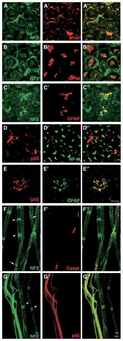
Figure 4.
Immunohistochemistry of merlin and p55 expressed in the neuronal tissues. Panels A–A″, rat nerve transverse section stained for NF2 and S100. Panels B–B″, rat nerve transverse section stained for NF2 and NF-M. Panels C–C″, rat nerve transverse section stained for NF2 and GFAP. Panels D–D″, rat nerve transverse section stained for p55 and NF-H. Panels E–E″, rat nerve transverse section stained for p55 with GFAP. Panels F–F″, mouse nerves teased fibers stained for NF2 and Caspr. Panels G–G″, mouse nerves teased fibers stained for NF2 and p55 using their respective antibodies as described in the text. Arrowheads indicate the location of Schmidt-Lanterman incisures in panels F and G. Arrows in panel F show the paranodes marked by Caspr. The right-hand panels represent the merged image of the two left-hand panels. Bar is 10 μm in A–G. A color version of this figure is available in the online journal.

