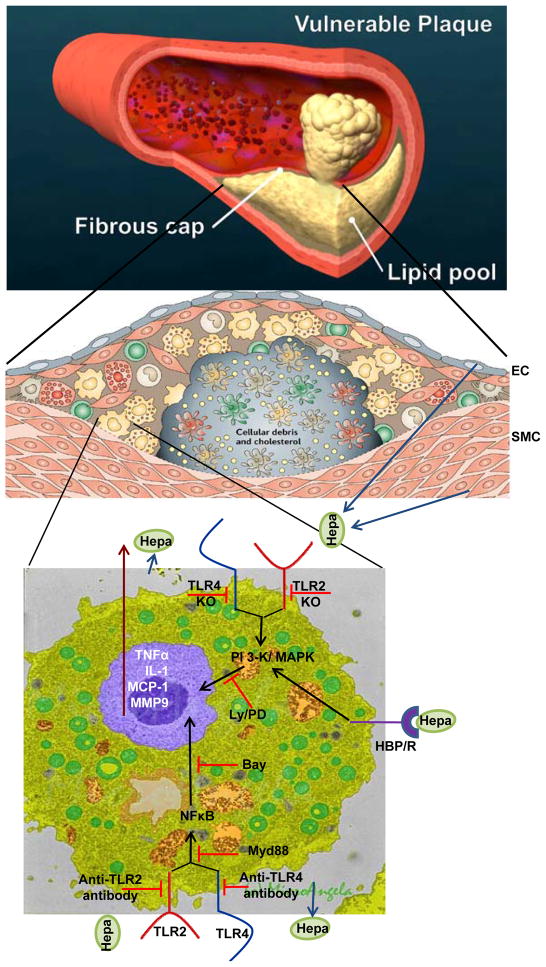
Figure 1.
Summary and proposed model for heparanase function in plaque vulnerability. A schematic illustration of blood vessel occluded with a vulnerable plaque is shown in the upper panel. A more detailed structure of the plaque is shown in the middle panel. A fibrous cap composed of endothelial cells (EC), smooth muscle cells (SMC) and collagen surrounds and stabilizes the plaque which is populated by macrophages, foam cells, monocytes, T cells and dendritic cells. Heparanase, secreted by EC, SMC or macrophages is abundantly present in the plaque and contributes to plaque rupture and vulnerability directly and indirectly. Heparanase activity, together with proteolytic activity exerted by MMP9 and other proteases weaken the fibrous cap making it more susceptible to rupture. Inactive heparanase activates macrophages and induces the expression of MMP9 and pro-inflammatory cytokines (i.e., TNF-α, MCP-1, IL-1), leading to the recruitment of more immune cells and plaque progression. Cytokine induction by heparanase involves the PI-3K and MAPK signaling pathways, NFκB, and TLR-2 and -4. The exact order of the complex sequence of events that lead to TLR activation and signaling cascade is not clear but is thought to be mediated by presently unidentified heparanase binding protein/receptor (HBP/R; lower panel).
Adopted from Hansson and Libby, Nature Immunology 6:508–519, 2006; and Microangela (http://www5.pbrc.hawaii.edu/microangela).

