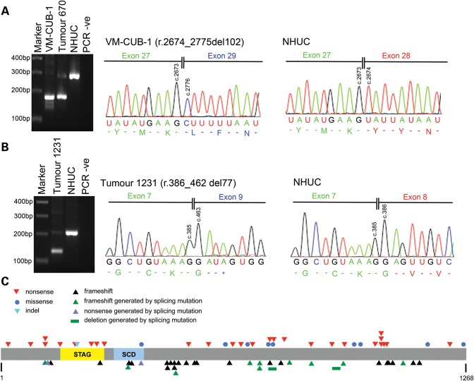
Figure 1.
STAG2 mutations in bladder cancer samples. STAG2 splice site mutations in (A) cell line VM-CUB-1 and (B) bladder tumour 1231. Left-hand panels in (A) and (B) show aberrantly sized bands generated by RT-PCR, and right-hand panels show nucleotide sequences of aberrant RT-PCR products illustrating the effects of splicing mutations at the RNA level. (A) PCR was performed on VM-CUB-1 cDNA using primers in exons 27 and 29. A PCR product 102 bp smaller than that generated using cDNA from normal human urothelial cells (NHUC) was observed for VM-CUB-1. Sanger sequencing of RT-PCR products showed that this aberrantly sized band lacked exon 28. Bladder tumour 670 carries a splice site mutation that is also predicted to result in skipping of exon 28, and this sample generated a PCR product similar in size to that observed for VM-CUB-1. Sequencing confirmed that this PCR product also lacked exon 28 (data not shown). (B) PCR was performed on cDNA from bladder tumour 1231 using primers in exons 7 and 9. A PCR product 77 bp smaller than that generated using cDNA from NHUC was observed for tumour 1231, and sequencing confirmed that the aberrantly sized PCR product lacked exon 8. A detailed description of splice site mutations in all samples is given in Supplementary Material, text. (C) Diagram of STAG2 protein showing position of somatic nonsense, missense, splicing, frameshift and indel mutations identified in bladder tumours. STAG and SMC (structural maintenance of chromosomes) domains are also shown.

