Figure 3.
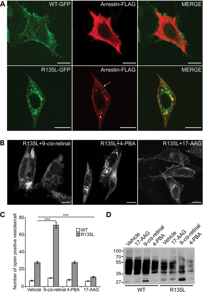
Pharmacological manipulation of R135L rhodopsin. (A) Subcellular distribution and trafficking of WT-GFP and R135L-GFP rod opsin (green) in SK-N-SH neuroblastoma cells co-transfected with visual arrestin-FLAG (red). WT rod opsin mainly decorated the PM and visual arrestin remained in the cytoplasm. R135L rod opsin mutant recruited and translocated cytosolic visual arrestin to the PM (arrow) and the endocytic compartments (arrowhead). Scale bars: 10 μm. (B) Fluorescence microscopy of R135L-GFP rod opsin and treated with 10 μm 9-cis-retinal, 10 mm 4-PBA and 1 μm 17-AAG. Scale bars: 10 μm. (C) Cell counts of intracellular vesicle incidence in cells expressing WT-GFP (open) or R135L-GFP (grey) following treatment with 9-cis-retinal, 4-PBA or 17-AAG for 18 h. Values are mean ± SEM. ***P < 0.0001, Student's t-test n = 3. (D) Western blot with 1D4 of untagged WT and R135L rod opsin after treatment with 9-cis-retinal, 4-PBA or 17-AAG for 24 h. The position of molecular-weight markers is indicated on the left in kDa.

