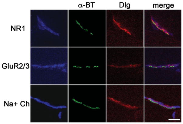
FIGURE 2.
Confocal coimmunofluorescence of AChRs (α-BT) (green) with Dlg (red), and NR1 or GluR2/3 (blue) show overlapping localization of Dlg and glutamate receptors at postsynaptic NMJs in rat. Dlg localization is also shown to overlap with Na+ Channels (blue), known to localize to secondary folds of the postsynaptic membrane. Merged images from the green, red, and blue channels are shown in the last column for each NMJ. Purple represents overlap of Dlg (red) with glutamate receptors (blue). Scale bar = 3.7 μm.

