
FIGURE 3.
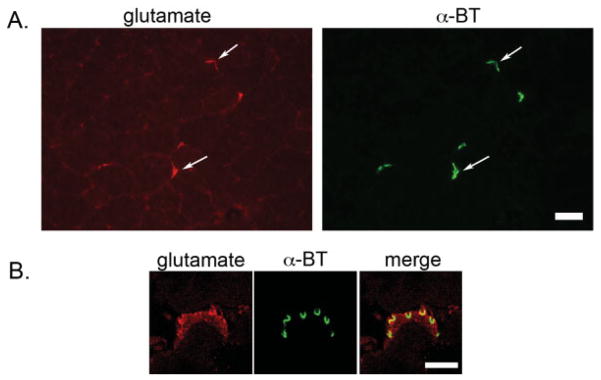
Localization of glutamate at mouse NMJs. A: Epifluorescence images of unfixed quadriceps sections shows localization of glutamate (red) specifically coincident with α-bungarotoxin-stained (α-BT) (green) AChRs at NMJs. Scale bar = 50 μm. B: Confocal images of fixed quadriceps sections show the subcellular localization of glutamate (red) with respect to AChRs (green) at the mammalian NMJ. Scale bar = 3.7 μm.