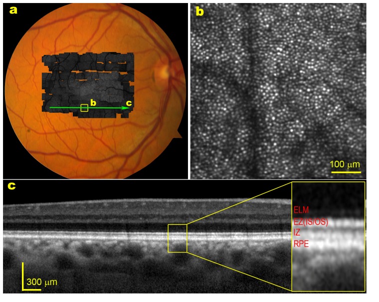
Fig. 1.
A healthy retina imaged by AO-SLO and SD-OCT. The subject is a 54-year-old man (white non-Hispanic). (a) The high resolution retinal image montage (gray image) taken with the AO-SLO is overlaid on color fundus photograph. (b) AO-SLO image contained in the box of panel a reveals clear photoreceptor mosaic. The bright spots are cones. The dark bands are the shadow of retinal capillaries. (c) SD-OCT taken through the green arrow-line in panel a.

