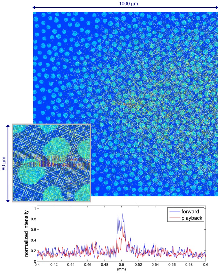
Fig. 9.
(Media 3 (2.2MB, MOV) ) (Top): Simulation of TRUE light propagating through a 1000-μm-by-1000-μm virtual tissue model consisting of ~670 cells. The 2-D virtual tissue model is constructed based upon a HT29 cell. (Bottom): The time-reversed light profile (playback scenario) is compared with the forward scenario light profile. Both field profiles were measured at the center of the simulation space. (Inset-figure): A 80-μm-by-80-μm zoomed-in view of back-propagated light converging at the virtual light source.

