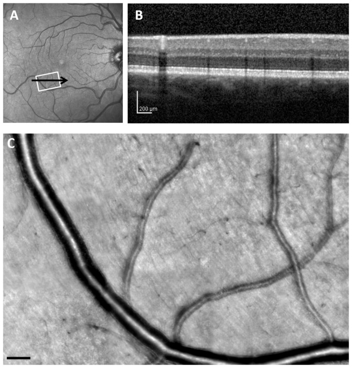
Fig. 1.
Retinal images from 35-year-old control subject (C 01), comparing a wide-field SLO en-face image, SDOCT cross section, and AOSLO high resolution montage. (A), 30 x 30 deg. wide-field SLO en-face image showing the region of interest. (B), SDOCT cross section, with the location as indicated by the black arrow in A. (C), Montage of AOSLO reflectance images, corresponding to the white rectangle in A. Scale bar = 100 μm.

