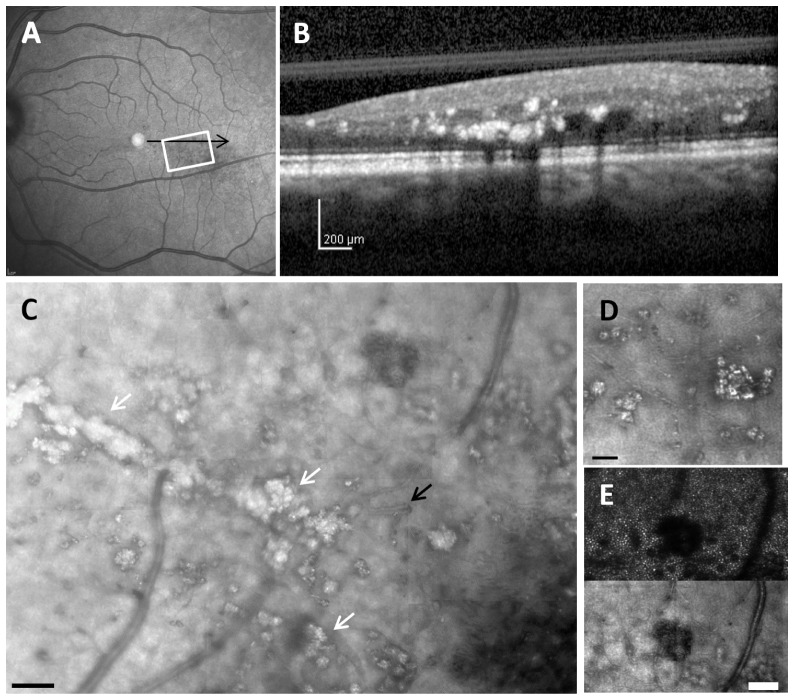
Fig. 4.
Retinal images from 47-year-old diabetic subject with mild NPDR and clinically significant macular edema. (A), 30 x 30 deg wide field SLO image showing hard exudates and edema. (B), SDOCT cross section, with the location as indicated by the black arrow in A, showing extensive hard exudates and intraretinal fluid. (C), montage of AOSLO reflectance image also showing corresponding extensive hard exudates (white arrows), and in addition surrounding numerous capillary abnormalities including vessel looping (black arrow). The dark region in the lower right corner is a typical appearance of loss of reflectivity due to the accumulation of fluid. Scale bar = 100 µm. (D), Enlargement of region of multiple hard exudates, showing sizes as small as 5 μm. (E), comparison of a region of edema as indicated by cysts in the inner retina, top- imaging focused on photoreceptors using a 2x Airy disc aperture, bottom- same region with offset aperture focused on blood vessels. For C scale bar = 100 μm, for D and E Scale bar = 50 μm.

