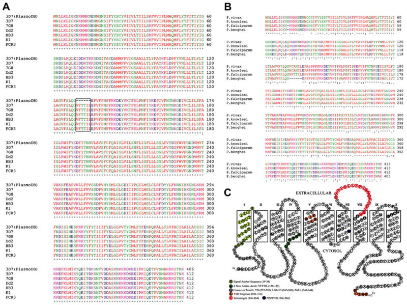
Fig. 1.
Parasite SPP sequences and topology model. (A) Alignment of 6 strains of PfSPP was performed using ClustalW2. The 3D7 sequence was taken from the PlasmoDB (Gene ID: PF14_0543). Only one amino acid difference was found in the FCR3 strain (180, A→S). (B) SPP alignment. (C) Topology model of PfSPP by ConPred II. Signal-anchor sequence (19–38); two active site motifs YD (227–228) and LGLGD (265–269); and the PALL (341–344) motif are indicated.

