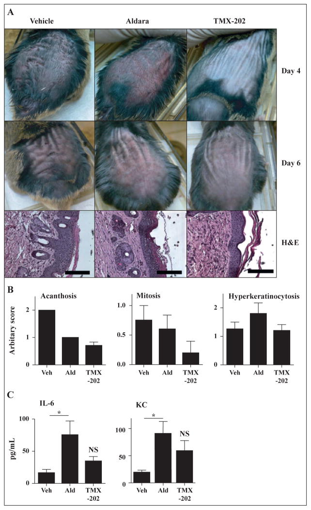
Figure 5. In vivo efficacy evaluation of TMX-202 gel.
The back skin of Inv-Myc mice (n = 5) was treated with 5mg/mL tamoxifen daily for days 0 to 7. Mice were treated with Aldara, TMX-202 or vehicle gel on days 0, 2, 4, 6, 8, and 10. (A and B) On days 4 and 6, the gross appearance of the skin was recorded (A). On day 11, the skin samples were collected and hematoxylin/eosin-stained samples were evaluated and scored for acanthosis, mitosis, or hyperkeratinocytosis (B). Data shown are representative of two independent experiments. (C) Levels of IL-6, and KC in sera samples 2 hrs after the first application. Data shown are mean ± SEM of pooled data of two independent experiments. * denotes p<0.05 by one way ANOVA with Bonferroni post hoc test.

