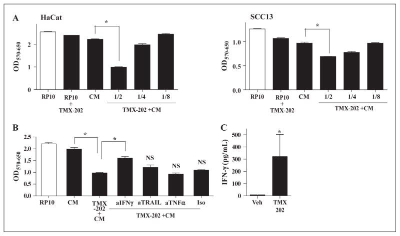
Figure 6. TMX-202 conditioned media suppressed the growth of keratinocytes.
Human PBMC (106/mL) were incubated with 1 μM TMX-202 in complete RPMI and the culture supernatants were used as conditioned media (TMX-202+CM). CM without TMX-202 or complete RP-10 were used as controls. (A) HaCaT or SCC13 cells were incubated with TMX-202 +CM at various dilutions (1/2, 1/4, or 1/8) for 48 hrs and the viability of cells was determined by MTT assay. (B) HaCaT cells were treated with 2 fold diluted media in the presence of antibodies against human IFN-γ, TRAIL or TNFα for 48 hrs. * denotes p<0.05 compared to the CM or TMX-202+ CM by one-way ANOVA with Bonferroni post hoc test. NS denotes “not significant” compared to TMX-202+CM. (C) PBMC from five independent donors were cultured with TMX-202, and IFN-γ in the culture supernatant was measured by Luminex beads assay. * denotes p<0.05 compared to vehicle treatment by Student t test.

