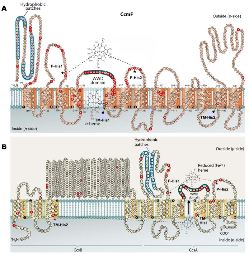
Fig. 4.
Topology of the system I CcmF protein from Escherichia coli (A) and the system II CcsBA fusion protein from Helicobacter hepaticus (B). Possible histidine axial ligands to heme are starred, and are designated P-His1, P-His2, TM-His1, and TM-His2. The highly conserved WWD domain and the hydrophobic patches are shaded. Completely conserved amino acids (red) were identified by individual protein alignments using CcmF or the CcsB and CcsA ORFs from selected organisms, as described in (Kranz et al., 2009). Diagram is modified from (Kranz et al., 2009).

