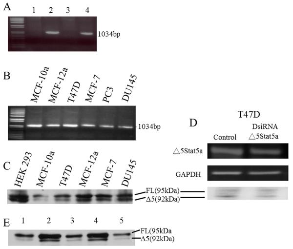
Figure 2. Expression of Δ5 Stat5a at mRNA and Protein Level.
A: Ethidium bromide stained gel showing the specificity of the primers for FL and Δ5 Stat5a. Lane 1, primers specific to FL used with plasmid for Δ5; lane 2, primers specific for FL used with plasmid for FL(amplicon of 1044bp); lane 3, primers specific for Δ5 used with plasmid for FL; lane 4, primers specific for Δ5 used with plasmid for Δ5 (amplicon of 1034bp). B: ethidium bromide stained gel showing Δ5 Stat5a-specific amplicons produced from cDNA in each cell type. C: Western blot showing both FL and Δ5 Stat5a protein in mostly the same cell lines as shown in B. The antibody used was against the C-terminus of Stat5a and therefore recognized both FL and Δ5 Stat5a protein. D: Effect of DsiRNA knockdown of Δ5 Stat5a on mRNA (upper panel) and protein (lowest panel). 5×10⁵ cells were seeded in 35mm wells. The next day, cells were transfected with 1μg DsiRNA or control. Cells were collected 24 hours after transfection and RNA/protein expression was analyzed by RT-PCR and Western blot. E: Western blot of FL and Δ5 Stat5a from five HUVEC cultures.

