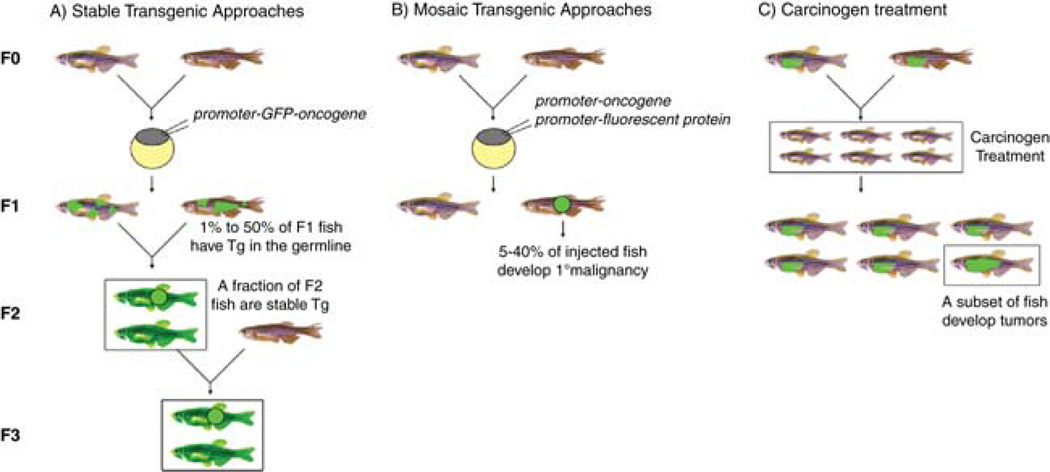
Fig. 1.
Methods for creating fluorescent-labeled cancers in zebrafish. Approaches for making stable (A) and mosaic (B) transgenic and chemically induced models (C) of fluorescent-labeled cancer. Stable transgenic animals capable of germline transmission are denoted by boxes inA. (C) F1 stable transgenic zebrafish treated as larva with carcinogen are denoted as smaller fish and contained within a boxed region. These animals are monitored for tumor onset, with a subset of animals developing fluorescent-labeled disease (denoted as an adult sized animalwith an enlarged region of GFP+ cells, single fish boxed). (See color plate.)

