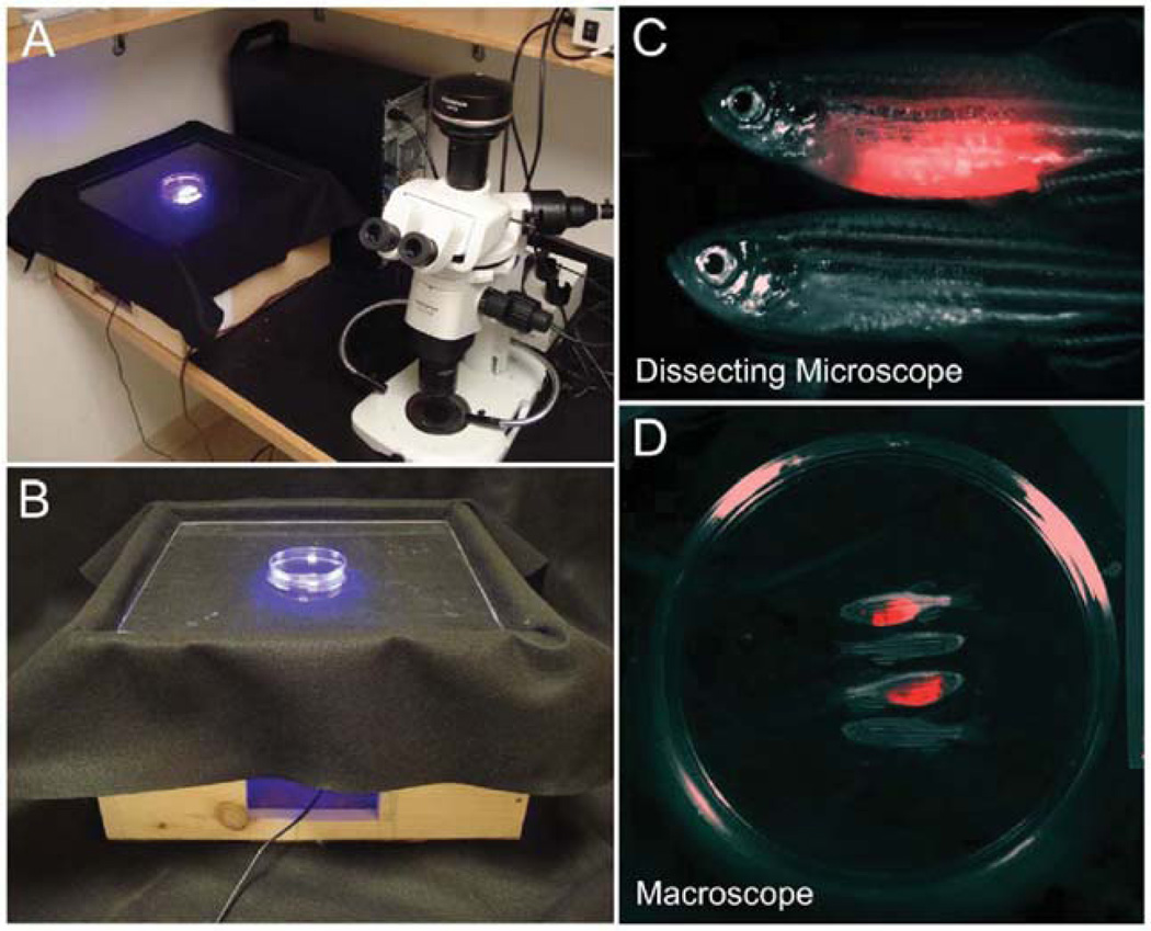
Fig. 2.
Macroscopic imaging of fluorescent tumor engraftment. (A) The LED fluorescence macroscope (left) juxtaposed with an epifluorescent Olympus SZ16 stereomicroscope (right). (B) Enlarged view of the LED fluorescence macroscope. (C) Images of two adult fish, one with a rag2-dsREDexpress+ T-ALL (top) and one control fish (bottom). (D) Macroscope image of four adult zebrafish two of which have developed rag2-dsREDexpress+ T-ALL. Petri dish is 10 cm in diameter.

