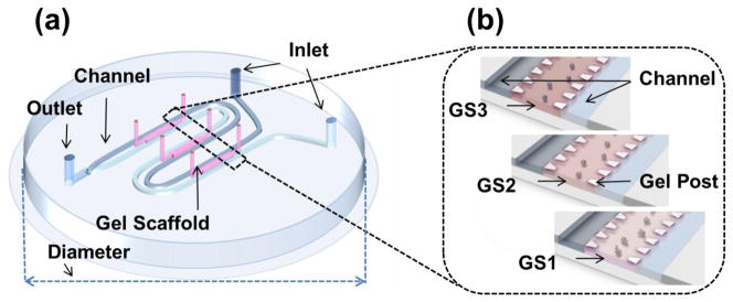
FIGURE 1.

General features of a simple 3D microfluidic platform for tracking microvasculogenic behaviors within differently engineered microenvironments. (a) Schematic representation of the microfluidic device. The device with an overall diameter of 34 mm consists of two independent flow channels with inlet dimensions of 500 μm (width) × 150 μm (height) and three GSs with dimensions of 4.1 mm (length) × 1.3 mm (width) × 150 μm (height). The two independent flow channels merge at the outlet. (b) Schematic representation of enlarged 3D GSs. Three 3D GSs were created by injecting three different concentrations of type-I collagen gel or three different compositions of collagen and/or fibrin gel mixed with HUVECs through the gel ports. The 3D GSs were designated as GS1 (top), GS2 (middle), and GS3 (bottom) according to their position on the device and on the direction of inlet. Each GS contained 20 trapezoidal posts that were used to confine the gel to the region between the rows of posts.
