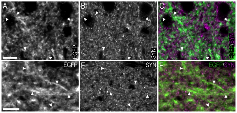
Figure 1. Double-immunofluorescence confocal micrographs showing the distribution of EGFP− and synaptophysin-LI in the bed nucleus of the stria terminalis and medial amygdaloid nucleus – posterodorsal part.
(A–C) Single-channel confocal micrographs of double-fluorescence immunohistochemistry (C), displaying EGFP− (A), and synaptophysin-LI (B) in the bed nucleus of the stria terminalis. (D–F) Single-channel confocal micrographs of double-fluorescence immunohistochemistry (F), displaying EGFP− (D), and synaptophysin-LI (E) in the medial amygdaloid nucleus – posterodorsal part. In A–F, arrowheads point to double-labelled EGFP-ir nerve fibres/terminals and synaptophysin-LI. Scale bars: A = 10 µm, applies A–C; D = 10 µm, applies D–F.

