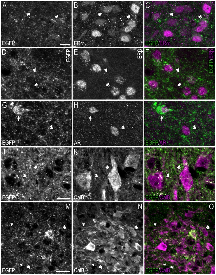
Figure 14. Double-immunofluorescence confocal micrographs showing apparent contacts between EGFP+ fibres/terminal-like structures, and ERα−, ERβ-, AR- or calbindin-ir cell bodies/fibres in the hypothalamus.
(A–C) Single-channel confocal micrographs of double-fluorescence immunohistochemistry (C), displaying EGFP− (A), and ERα-LI (B). The confocal micrographs in (A–C) are magnified views of the immunohistochemistry presented in Fig. 9A–F. (D–F) Single-channel confocal micrographs of double-fluorescence immunohistochemistry (F), displaying EGFP− (D), and ERβ-LI (E). Confocal micrographs in (D–F) are magnified views of Fig. 9G-L. (G–I) Single-channel confocal micrographs of double-fluorescence immunohistochemistry (I), displaying EGFP− (G), and AR-LI (H). Confocal micrographs in (G–I) are magnified views of Fig. 9M-R. (J–L) Single-channel confocal micrographs of double-fluorescence immunohistochemistry (L), displaying EGFP− (J), and calbindin-LI (K). (M–O) Single-channel confocal micrographs of double-fluorescence immunohistochemistry (O), displaying EGFP− (M), and calbindin-LI (N). In A–O, arrows point to double-labelled cell bodies, arrowheads point to apparent contacts or close anatomical association between EGFP-ir nerve fibres and calbindin-ir fibres (O), and double arrowheads point to apparent contacts or close anatomical association between EGFP-ir nerve fibres and single-labelled ERα− (C), ERβ- (F), AR-ir (I) or calbindin-ir (L, O) cell bodies. Scale bars: A = 10 µm, applies A–I; J = 10 µm, applies J–L; M = 20 µm, applies M–O.

