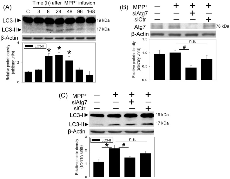
Figure 2. MPP+ induced autophagy in the nigrostriatal dopaminergic system of rat brain.
Intranigral infusion of MPP+ (3 µg/µl) was performed in the anesthetized rats. Rats were sacrificed at the indicated times. (A) Time-dependent effects of MPP+ on LC3-II level in the substantia nigra (SN) were studied using Western blot assay. Graphs show statistical results from relative optical density of bands on the blots estimated by Image J software. Values are the mean±S.E.M. (n = 3/group). *P<0.05 in the MPP+-treated groups compared with the control groups by one-way analysis of variance (one-way ANOVA) and followed by the LSD test as post-hoc method. (B and C) Intranigral infusion of Atg7siRNA was performed 4 days prior to intranigral infusion of MPP+ (3 µg/µl) in chloral hydrate-anesthetized rats. Rats were sacrificed 30 h after intranigral infusion of MPP+. The levels of Atg7protein (B) and LC3-II (C) levels in SN were measured using Western blot assay. Each lane contained 25 µg protein for the experiments. Graphs show statistical results from relative optical density of bands on the blots estimated by Image J software. Values are the mean±S.E.M. (n = 4/group). *P<0.05 in the MPP+-infused SN compared with the intact SN, # P<0.05 in Atg7siRNA transfected and MPP+-infused SN compared with MPP+-infused SN, n.s. for not significant by Kruskal-Wallis test and followed by Mann-Whitney U test as post-hoc method.

