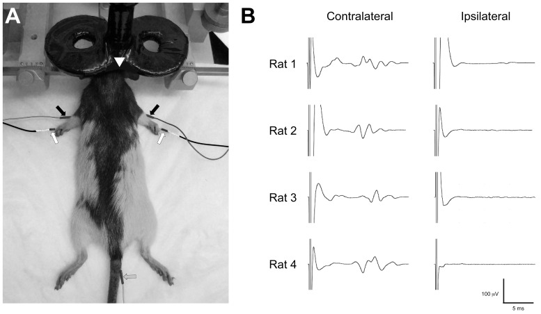
Figure 1.
(A) EMG-rTMS rat setup. Photograph shows rat in stereotaxic frame. Monopolar stainless steel needle electrodes are placed into the brachioradialis of each forelimb and between the third and fourth digit in the footpad (arrows). A ground electrode is inserted in the tail. A 40 mm figure-of-eight coil is fixed to a micromanipulator arm and positioned over the left or right hemisphere. (B) Demonstration of contralateral activation by TMS in rat. Representative brachioradialis MEPs (average of ten consecutive sweeps) are shown. Note, at motor threshold, lateralized TMS elicits isolated MEP in the contralateral forelimb.

