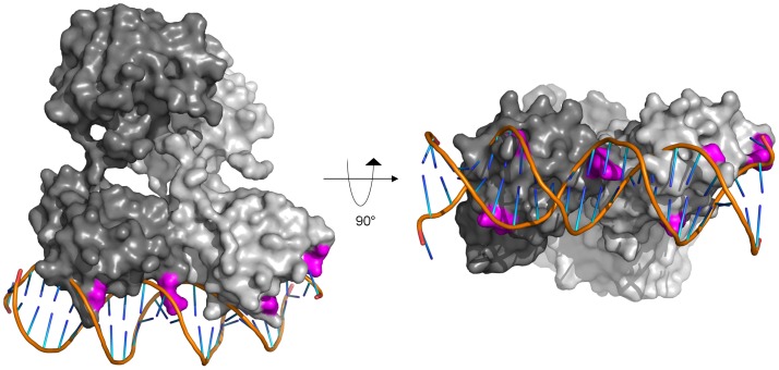
Figure 9. Proposed model of full-length ChxR binding to tandem DNA repeat.
The solution state of full-length, dimeric ChxR (each polypeptide colored a different shade of gray, surface representation) was manually overlayed onto the cocrystal structure of the PhoB effector domain from E. coli bound to its cognate pho box (PDB ID: 1GXP). The structure is rotated 90° about the protein:DNA interface on the right. Surface exposed residues within ChxR that were implicated in DNA binding by site-directed mutagenesis are highlighted in magenta.

