
Figure 6. Chloroquine enhances FTS-induced cell death.
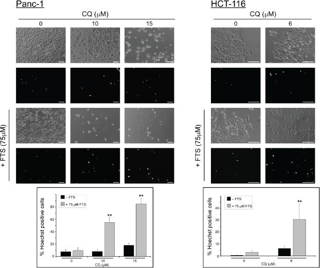
Panc-1 and HCT-116 cells were treated with 75 µM FTS, with or without chloroquine, at the indicated concentrations for 7 or 6 days, respectively. The cells were stained with the fluorescent DNA dye bisbenzimide (Hoechst 33258, 1 µg/ml) to assess the number of dying cells. Following staining, the cells were photographed using Olympus motorized inverted research microscope Model IX81 (20×magnifcation for HCT-116 cells and 10×magnifcation for Panc-1 cells ; scale bars, 100 micrometer). Upper panel, representative images. Lower panel, percentage of dying cells was estimated by counting the number of Hoechst-positive cells compared to the number of total cells in each field (7-10 fields for each treatment, 100-200 cells per field). Results are presented as mean ± S.D (**, p < 0.01, compared to each treatment alone).