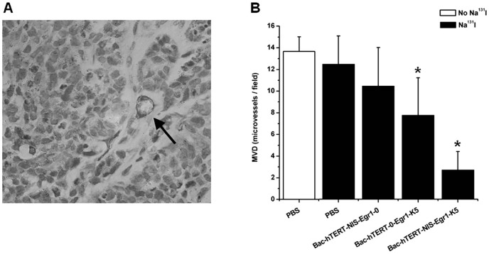
Figure 7. Immunohistochemical staining of CD31 and MVD determination.
(A) Immunohistochemical staining with anti-CD31 antibody. The arrow indicates a representative microvessel within the tumor (magnification, 400×). (B) MVD was determined by identifying the three most intense areas of neovascularization of each sample and counting at low power (magnification, 100×). Average vessel counts of the three selected fields were recorded.*P<0.05.

