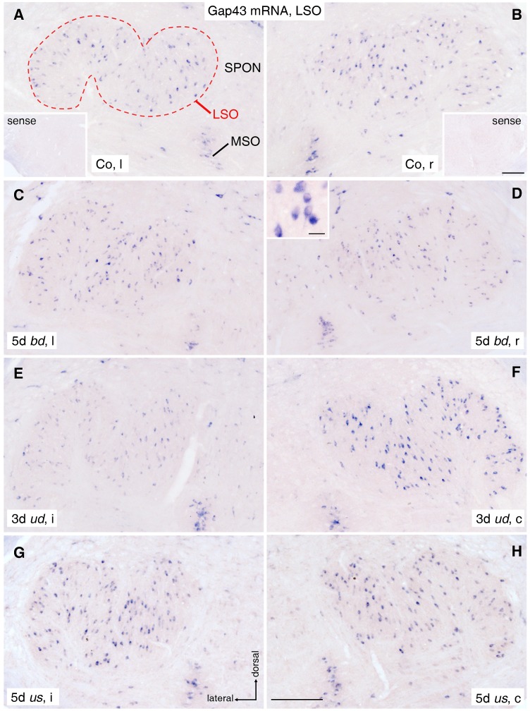
Figure 4. Gap43 mRNA staining in lateral superior olive (LSO).
(A, B) A basal level of Gap43 mRNA was seen in neuronal cell bodies (purple dots) of the entire left (A) and right (B) LSO (dashed outline) of untreated control rats (Co). The insets show lack of staining in corresponding sections after use of Gap43 sense probe. Scale bar for insets: 0.2 mm. (C, D) Gap43 mRNA expression in left (C) and right (D) LSO after 5 days (d) of bilaterally deaf (bd) rats is equivalent to control level. Inset in D shows Gap43 mRNA positive neurons at higher magnification. Scale bar of inset: 20 μm. (E) Following 3d of unilateral deafness (ud), Gap43 mRNA expression decreased in neurons of LSOi compared to controls. (F) Simultaneously, the expression increased contralaterally. (G, H) After 5d of unilateral stimulation (us), high bilaterally balanced Gap43 mRNA levels were observed. Scale bar for A to H: 0.2 mm. SPON: superior paraolivary nucleus; MSO: medial superior olive; l: left; r: right; i: ipsilateral; c: contralateral.

