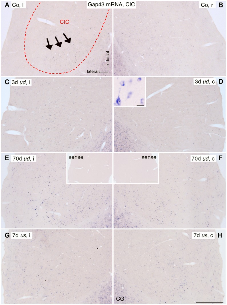
Figure 5. Gap43 mRNA staining in central inferior colliculus (CIC).
(A, B) In the untreated control group (Co), Gap43 mRNA positive neurons (purple dots) in CIC (dashed outline) were mainly localized ventrally (arrows). (C, D) 3 days (d) after unilateral deafness (ud), Gap43 mRNA level increased in CICi (C), while expression was decreased in CICc (D). The inset shows a higher magnification of stained CIC neurons. Scale bar in inset: 20 μm. (E, F) After 70d of ud, Gap43 mRNA expression rose bilaterally in CIC, with a significantly higher level on the ipsilateral side (E) compared to the contralateral side (F). Insets show control staining after incubation with Gap43 sense probe. Scale bar for insets: 0.2 mm. (G, H) Unilateral stimulation (us) for 7d resulted in an increase of Gap43 mRNA levels in ventral CIC on ipsilateral (G) and contralateral (H) side. Scale bar for A to H: 0.2 mm. l: left; r: right; i: ipsilateral; c: contralateral; CG: central gray.

