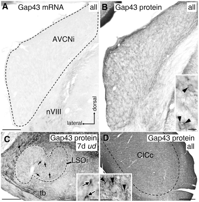
Figure 8. Gap43 mRNA and protein expression in auditory brainstem.
(A) Anteroventral cochlear nucleus (AVCN, dashed line) was devoid of Gap43 mRNA staining on both sides under any experimental condition. (B) Faintly stained Gap43 protein-positive axonal boutons were present throughout AVCN in controls and all experimental conditions. Inset: higher magnification of immuno-positive presynaptic endings (arrowheads). (C) Gap43 protein expression in lateral olivocochlear neurons (arrows) within LSOi (dashed line) required at least 5 days (d) of electrode implantation independent of its activation. Inset: close-up of Gap43 protein-positive neurons (arrows) and boutons following 7d of ud. (D) Throughout CIC (dashed line), Gap43 immunoreactivity was always present. Inset: close-up of immuno-positive presynaptic endings (arrowheads). Scale bars for A to D: 0.2 mm. Scale bars of insets B to D: 20 μm. LSO: lateral superior olive; CIC: central inferior colliculus; nVIII: 8th cranial nerve; tb: trapezoid body; i: ipsilateral; c: contralateral; ud: unilateral deafness.

