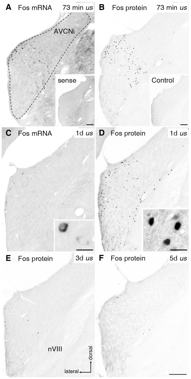
Figure 9. The effectiveness of electrical intracochlear stimulation (EIS) on neurons of the anteroventral cochlear nucleus (AVCN).

Fos mRNA (A) and Fos protein (B) closely corresponded locally after EIS for under 2 h duration. (A, C) Fos mRNA expression (black dots) in neurons of ipsilateral (i) AVCN after 73 min (A; cp. Illing and Rosskothen-Kuhl, 2012) and 1 day (d) (C) of unilateral stimulation (us). Fos-positive neurons were spatially limited to regions tonotopically corresponding to frequencies processed at the site of intracochlear electrode position. The number of labeled neurons was higher after 73 min than after 1d of us. Inset A: AVCNi was devoid of staining after incubation with Fos sense probe. Inset C: higher magnification of mRNA-positive neurons, showing strongest staining in cytoplasm. (B, D) Fos protein staining (black dots) in AVCNi after 73 min (B; cp. Illing and Rosskothen-Kuhl, 2012) and 1d (D) of us. Protein expression was spatially limited to a band tonotopically corresponding to intracochlear stimulation position. Inset B: AVCNi of control. Scale bars in insets A and B: 0.2 mm. Inset D: higher magnification of Fos protein positive nuclei in AVCNi. Scale bars in insets C and D: 20 μm. (E) Three days after sustained EIS a strong decrease in the number of Fos protein positive nuclei was observable. (F) Following 5d of us, no further protein positive nuclei were detectable. Scale bar for A to E: 0.2 mm. nVIII: 8th cranial nerve.
