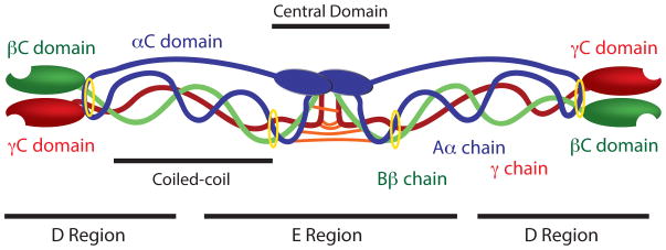
Figure 1. Fibrinogen Structure.
Aα chains are shown in blue, Bβ chains are shown in green, and γ chains are shown in red. Interchain disulfide bridges connecting the six polypeptide chains in the central domain are shown in orange and disulfide rings stabilizing the coiled coil regions are shown in yellow.

