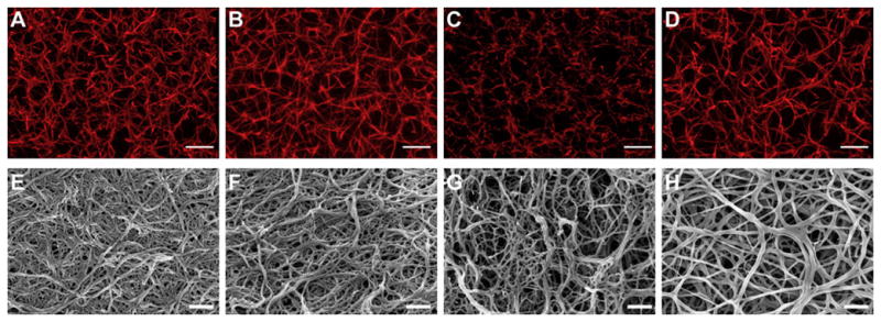
Figure 6. Confocal images of clots formed in the presence of synthetic B knob mimic AHRPYAAC conjugated to 5 kDa PEG.

Confocal and scanning electron microscopy: Representative confocal micrograph projections (10 μm z-stacks; A–D) and SEM images (E–H) from no peptide control (A and E), control peptide conjugate, GPSPFPAC-PEG (B and F), knob ‘A’ conjugate, GPRPFPAC-PEG (C and G), and knob ‘B’ conjugate, AHRPYAAC-PEG (D and H). Confocal: Objective = 63×, Scale bar = 10 μm. SEM: Magnification = 50,000×, Voltage = 3.0 kV, Scale bar = 500 nm. From Stabenfeldt SE, Gourley M, Krishnan L, Hoying JB, Barker TH: Engineering fibrin polymers through engagement of alternative polymerization mechanisms, Biomaterials, Copyright © 2012 by Elsevier. Reprinted by permission of Elsevier.
