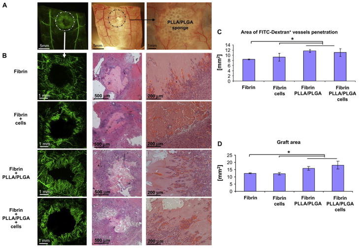
Figure 7. Fibrin PLLA/PLGA composite gels facilitate graft neovascularization and perfusion.
(A) Low power images of FITC-Dextran (green) and bright-field images highlighting the graft area. (B) Zoom at the graft area revealed intense FITC-Dextran+ neovessels penetration following 10 days postimplantation (left). Histological H&E examination demonstrated neovessels occupied with red blood cells penetrating into the graft area. Quantification of FITC-Dextran+ vessel area penetrating into the graft area (C) as well as calculation of the graft area size (D) revealed that Fibrin + PLLA/PLGA (with or without cells) constructs were most supportive of graft neovascularization and perfusion (n = 3–6). From Lesman A, Koffler J, Atlas R, Blinder YJ, Kam Z, Levenberg S: Engineering vessel-like networks within multicellular fibrin-based constructs, Biomaterials, Copyright © 2011 by Elsevier. Reprinted by permission of Elsevier.

