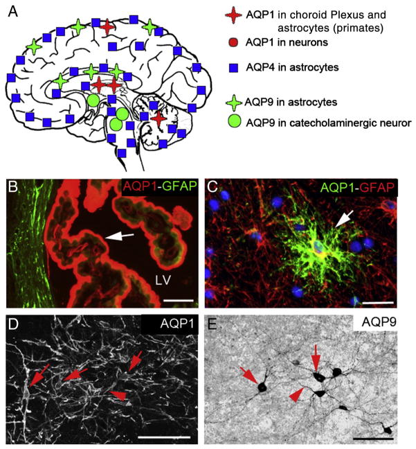
Fig. 1.
Aquaporin 1, 4 and 9 distributions in the brain. (A) Schematic drawing of aquaporin 1, 4 and 9 distributions in the brain. AQP1 is mainly observed in the choroid plexus and in some neurons. AQP1 is present in some astrocyte in primates. AQP4 is present in astrocytes in all brain structures with different levels and patterns of expression (see Fig. 2). AQP9 is mostly present in astrocytes and catecholaminergic neurons. (B) AQP1 immunostaining (red) in choroid plexus epithelium (arrow) located in the lateral ventricle (LV). The border of the LV is outlined by the GFAP staining (green), specific marker of the astrocytes. (C) AQP1 staining (green) in monkey cerebellum is co-localized (arrow) with GFAP staining (in red), suggesting the presence of AQP1 in a sub-population of astrocytes in primates. (D) AQP1 labeling is present in some neurons of the septum in the rat brain. (E) AQP9 immunoreactivity in catecholaminergic neurons of the ventral tegmental area. Bars: B, D = 50 μm; C, D = 40 μm.

