Fig. 2.
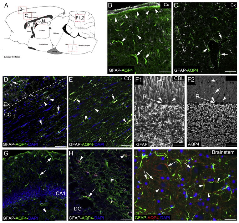
Variety in astrocyte AQP4 distribution. (A) Sagittal drawing of rat brain with the location of the pictures of AQP4 distribution in different brain areas. (B) AQP4 labeling (green) in the parietal cortex (Cx) is abundant on the glia limitans (arrow heads), revealed in gray by the GFAP staining. The AQP4 labeling is underlining the blood vessels (arrows) by its concentration in the astrocyte endfeet stained by the anti-GFAP (gray). (C) AQP4 distribution in the deeper cortex layer showed the “polarization” of the AQP4 labeling around the blood vessels (arrows). The double staining of AQP4 and GFAP exhibits its presence on the astrocyte endfeet (arrows). (D) AQP4 staining (green) in the corpus callosum (CC) is abundant around the blood vessel (arrows) and at distance from the perivascular space (arrowheads). In the white matter structure, AQP4 exhibits a different pattern of staining, with distribution of the protein on the astrocyte processes (gray, GFAP staining), possibly in association with node of Ranvier. (E) AQP4 staining (green) at higher magnification in the corpus callosum (CC) shows the co-localization with GFAP staining (gray, arrows and arrowheads). In contrast with the Cx, the AQP4 staining is spread in all the structure with “patchy” distribution, following the direction of the neuronal processes. (F1, F2) GFAP (F1) and AQP4 (F2) staining in the cerebellum differs from the cortex, with an abundant staining around all the neurons of the granular layer as well as around the Purkinje cell bodies (P, arrowheads). AQP4 is observed around the blood vessels in the molecular layer, where the radial glia is located. (G) AQP4 labeling (green) in the location of CA1 of the hippocampus outlines the blood vessels but it is also present in astrocyte processes in the stratum radiatum layer. (H) AQP4 labeling (green) in proximity of the dentate gyrus (DG) in the hippocampus. AQP4 is around the blood vessels (arrow) and in astrocyte processes remote of the perivascular space (arrowheads). (I) AQP4 labeling (red) shows staining around the neuronal cell bodies (arrows) as well as in contact with blood vessels (arrowheads). AQP4 (red) is co-localized with the GFAP staining (green). Bars: B, C, D, E, H = 50 μm; F1, F2 = 40 μm; G = 100 μm; I = 25 μm.

