Fig. 3.
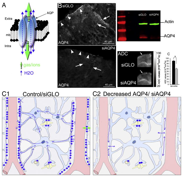
AQP4 water diffusion in the brain. (A) Schematic drawing of the aquaporin homo-tetramer assembly within the lipid membrane resulting in a central pore permeable to cations and gases (green arrows). Each individual aquaporin facilitates bi-directional water movement depending on the osmotic gradient (blue arrows, adapted from [33]). (B) AQP4 labeling in siGLO and siAQP4 treated rats showed a significant decrease in the intensity of AQP4 staining in the glia limitans and perivascular astrocyte in the cortex of siAQP4 treated rats. AQP4 Western blot (red) showed a significant decrease of the intensity in siAQP4 compared to siGLO treated rats. Decrease of the AQP4 is associated with a decrease of the water diffusion. Enlarged ADC images focusing on the contralateral cortex showed the decrease in ADC in siAQP4 compared to siGLO treated rats. The quantification showed a 50% decrease of the ADC signals where the AQP4 expression is decreased by 30% (modified from [35]). (C) Schema depicting the location of the APQ4 in brain cortex in control/siGLO condition (C1) and after siAQP4 injection (C2). The presence of AQP4 facilitates the water movement in the astrocyte network (C1), and after silencing APQ4 (C2), lower ADC values are caused by decreased water permeability due to a decreased number of AQP4 channels in perivascular space (C2).

