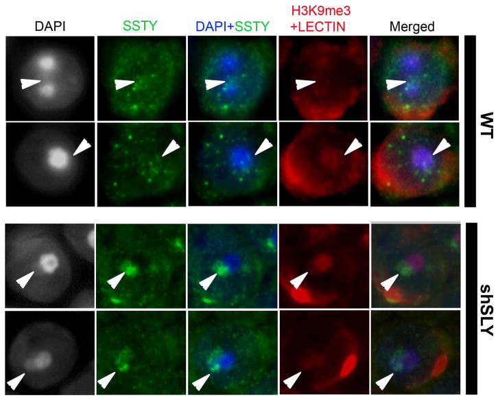
Figure 3. SSTY proteins colocalize with the PMSC and associated epigenetic marks.
Representative pictures of immunodetection of SSTY proteins (green) in the nucleus of WT and shSLY spermatids, taken at a high magnification (x100). In this immunodetection protocol, a permeabilization step was added prior to blocking and incubation with the primary antibody. DAPI (grey or blue) was used to stain nuclei. Lectin-PNA (red) was used to stain acrosomes for staging purposes. The PMSC (indicated by white arrowheads) can be visualized by DAPI staining (grey or blue) and H3K9me3 (red) staining (The most densely stained structure is the chromocenter; the structure less stained near the chromocenter is the PMSC). In WT spermatids, SSTY proteins are visible as bright foci, some of which colocalize with the PMSC. In shSLY spermatids, SSTY protein signal clearly colocalizes with the PMSC.

