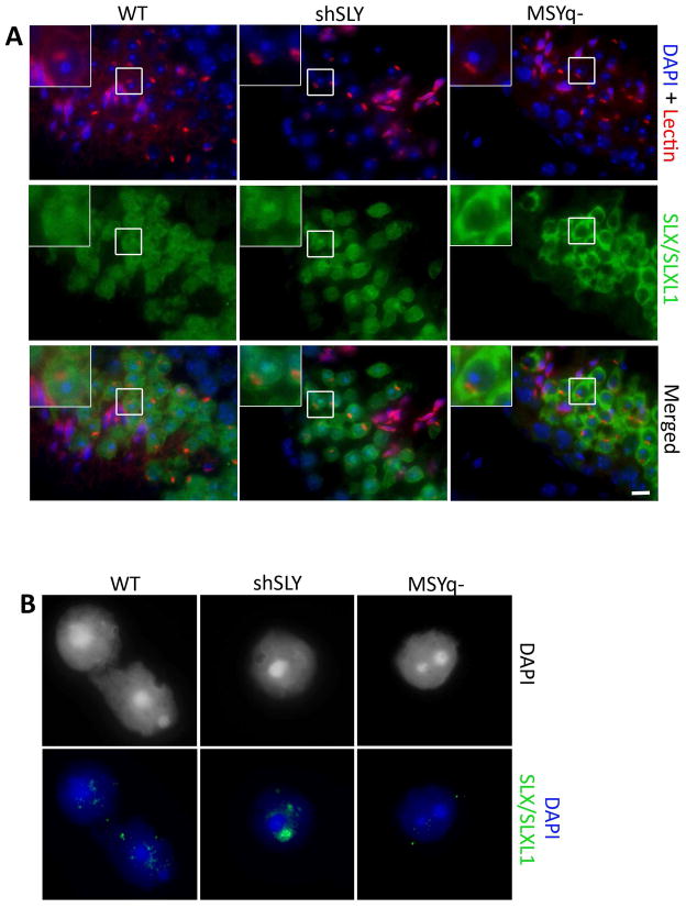
Figure 5. SLX/SLXL1 proteins are not visible in the nucleus of MSYq− spermatids.
(A) Detection of SLX/SLXL1 proteins (green) by immunofluorescence in WT, shSLY and MSYq− testis sections. DAPI (blue) was used to stain nuclei and lectin-PNA (red) was used to stain acrosomes. The inset in the upper left corner represents a 2.5 magnification. Scale bar indicates 10μm. (B) Representative pictures of the detection of SLX/SLXL1 proteins (green) by immunofluorescence in WT, shSLY and MSYq− round spermatid nuclei (surface spread technique). DAPI (grey or blue) was used to stain nuclei. No signal can be detected in MSYq− round spermatid nuclei.

