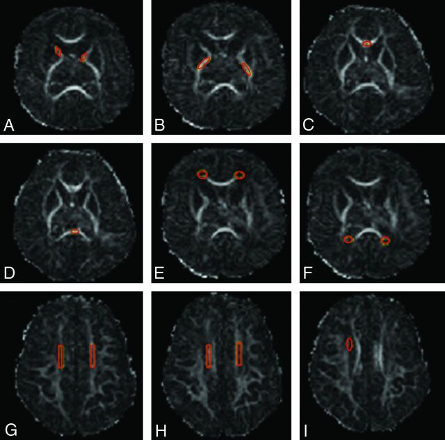
Fig 1.
Region-of-interest templates and placements shown on FA maps. A, Anterior limb of the internal capsule, B, Posterior limb of the internal capsule. C and D, Corpus callosum, genu and splenium. E, Frontal periventricular zone. F, Occipital periventricular zone. G and H, Centrum semiovale at 2 consecutive levels. I, Subventricular zone (right-sided). The same templates were used for all scans.

