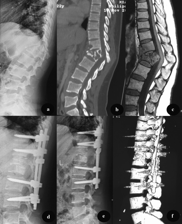
Fig. 2.
A pre-operative X-ray of a 45-year-old male demonstrated the destruction of T12 and L1, with a kyphosis angle of 43°. CT and MRI (b, c) show vertebral bone destruction and paravertebral abscess formation, severed spinal cord compression. The patient underwent one-staged anterior debridement, autologous bone grafting and posterior instrumentation. d A lateral X-ray indicates that kyphosis was corrected to 6° 3 months after surgery. e, f At 18-month follow-up, fixation was in good shape, without signs of tuberculosis recurrence

