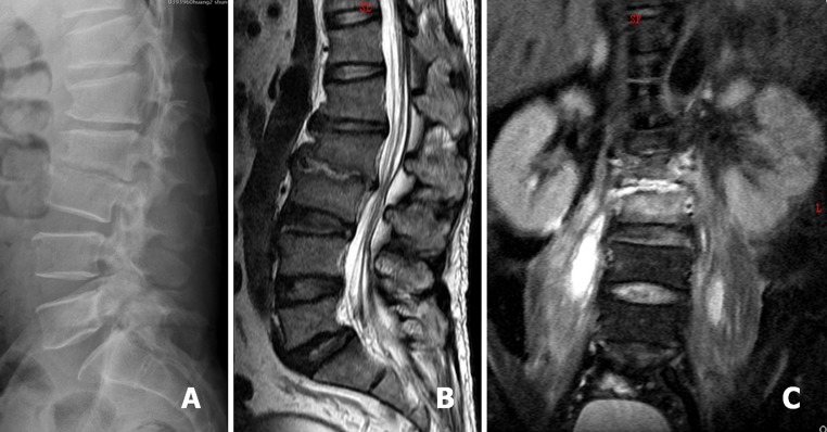
Fig. 1.

A 62-year-old man was diagnosed as L2–3 infectious spondylitis. Lateral radiograph showed L2–3 disc space collapse and endplates erosion (a). Sagittal and coronal T2-weighted magnetic resonance imaging (MRI) demonstrated a L2–3 infection source and associated bilateral psoas muscle abscess (b, c)
