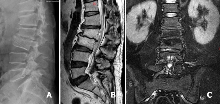
Fig. 2.

After PELD treatment, negative-pressure Hemovac with two drainage tubes was inserted into the L2–3 disc space for further continuous drainage of the offending pathogens (a). Sagittal T2-weighted MRI revealed L2–3 disc space narrowing, proceeding to spontaneous fusion (b). Coronal T2-weighted MRI demonstrated the abscess had disappeared at postoperative 8 months (c)
