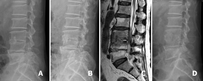
Fig. 3.

A 60-year-old man was diagnosed as having L3–4 infectious spondylitis and underwent L3–4 debridement and associated anterior interbody fusion with anterior iliac autograft. The lateral radiograph showed the bone graft located at the L3–4 anterior disc space (a). L3–4 recurrent infection developed in this patient and the follow-up lateral radiograph showed L3–4 posterior endplates erosion (b). Sagittal T2-weighted MRI revealed L3–4 infection with pus accumulation at the posterior disc space (c). The postoperative lateral radiograph showed the drainage tube inserted into the L3–4 posterior disc space for continuous drainage after PELD treatment (d)
