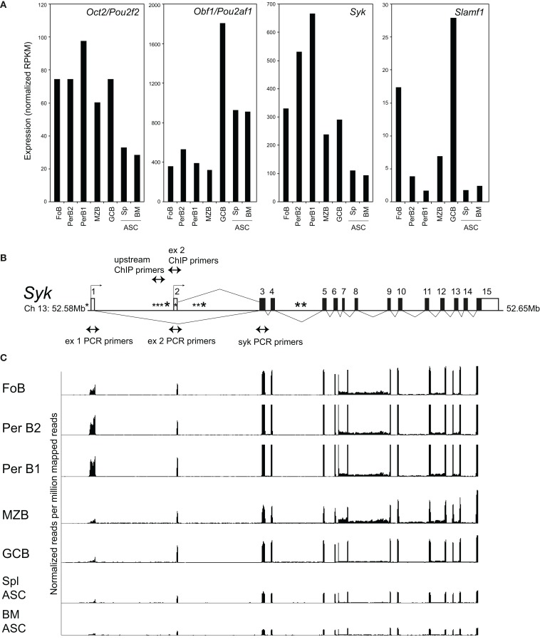
Figure 1.
Expression of Oct2, Obf1, Syk, and Slamf1 in peripheral B cell populations. (A) RNAseq data measuring expression of Oct2/Pou2f2, Obf1/Pou2af1, Syk, and Slamf1 in B cell populations sorted ex vivo from naïve C57BL/6 mice. FoB, follicular B cells from spleen (small B220+, IgM+, IgD+) PerB1 and PerB2, B220+ cells from peritoneal lavages of naïve mice, stained with CD23 and Mac1. B1 cells were CD23− and Mac1lo and B2 cells were CD23+ and Mac1−; MZB, splenic marginal zone B cells, B220+, IgMhi, CD21hi; GCB, germinal center B cells (B220+, Fas+, GL7+) from spleens of mice immunized 8 days previously with SRBC; ASC, antibody-secreting cells sorted as syndecan1+, GFP+ cells from spleens (Spl), and bone marrows (BM) of mice carrying the Blimp-GFP reporter gene (47). Data were derived from at least two independent biological replicates in all cases. Because Ig sequences can represent >70% of the RNA from plasma cells (data not shown), the RNAseq data shown in the figure excludes all reads mapping to the Ig (heavy and light chain) loci as described in Section “Materials and Methods.” (B) Structure of the mouse Syk gene, showing exons, alternative transcriptional start sites (small arrows), the locations of a perfect consensus octamer motif (*) and the positions of PCR primers used here. Filled boxes indicate protein coding sequence, and open boxes, sequence comprising the 5′ and 3′ untranslated regions of Syk mRNA. (C) RNAseq tracks showing expression of the Syk gene exons in different sorted B cell populations, normalized to library size, and aligned with the gene structure of (B). Note that exon 1, as shown in this panel, is not included in the RefSeq (Mouse mm9, July 2007) map of Syk mRNA, but is represented in alternate Syk transcripts ENSMUST00000120135 and ENSMUST00000118756 in the Ensembl database.

