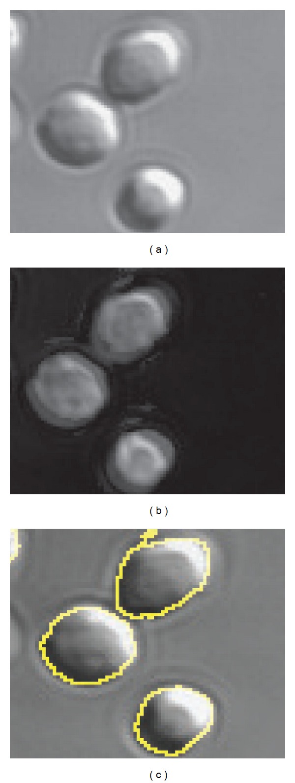
Figure 7.

(a) DIC images of Methanosarcina acetivorans grown on methanol. (b) After applying Hilbert transform to DIC images and adding them to the original image, the boundaries of the cells become clearer. (c) CellProfiler is used to perform segmentation on images in (b). Here identified cell boundaries are superimposed onto original DIC image to illustrate segmentation.
