Fig 1.
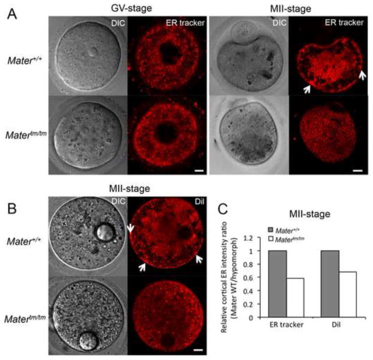
Altered endoplasmic reticulum localization and decreased cortical ER clustering in metaphase II-arrested Matertm/tm oocytes. (A) GV stage and MII stage Mater+/+ and Matertm/tm oocytes were stained with ER tracker and imaged using confocal microscopy. (B) DiI was microinjected into mature MII-arrested Mater+/+ and Matertm/tm oocytes and the localization of DiI (an ER marker) was documented using confocal microscopy. DIC images of microinjected oocytes highlight the DiI-containing oil-drop. Arrows highlight the cortical ER clusters in Mater+/+ oocytes, which were markedly reduced in Matertm/tm oocytes. Scale bar: 10 μm. (C) Semi-quantitative analysis of the cortical ER immunofluorescence signals (relative intensity) with an arbitrary number of the intensity ratio of Mater+/+ MII oocytes compared to Matertm/tm MII oocytes using Image J software.

