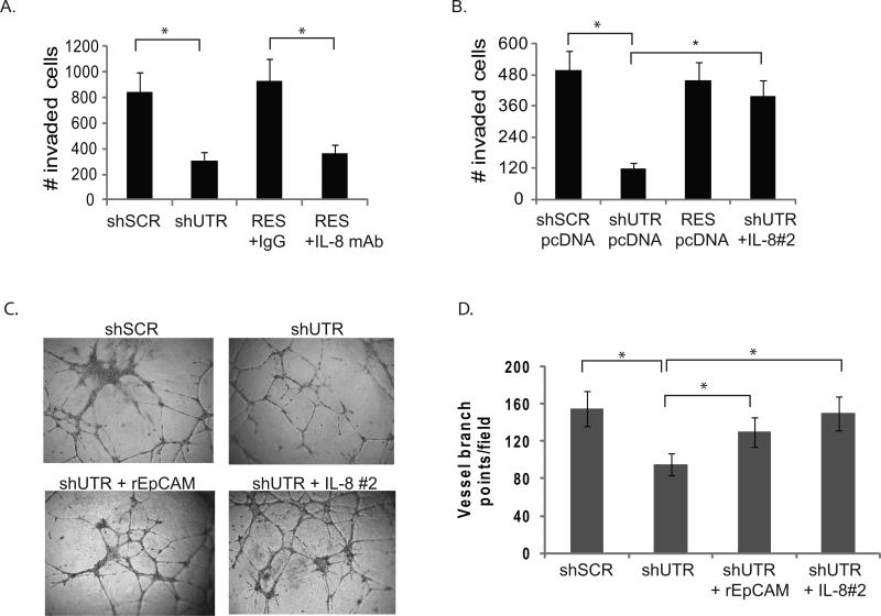
Figure 3. EpCAM-dependent modulation of IL-8 expression contributes to breast cancer invasion and angiogenesis.
(A-B) MDA-231 cells were stably transduced with lentiviral shRNA constructs targeting EpCAM (shUTR), or an irrelevant nucleotide sequence (shSCR). Transduced MDA-231 cells were subsequently rescued with vector (shUTR, see Figure S2). Trypsinized cells were washed with 10% FBS and Opti-MEM (Invitrogen). Mouse IgG or IL-8 neutralizing antibody (R&D systems, clone 6217.111) was added to cells, and the cells were transferred to matrigel chambers. Triplicate sets were harvested after 24h stained and counted. B. In independent experiments, tumor cell invasion was assessed using MDA-231 cells containing, shSCR, shUTR, shIL-8, EpCAM resistant to RNA interference (RES), or with a genetic construct encoding IL-8 (two clones were analyzed independently, designated IL-8#1, IL-8#2 see Figure S4). Invasion was evaluated in a matrigel transwell invasion assay. (C, D) MDA-231 cells were stably transduced with the lentiviral vectors as detailed above. EpCAM signaling was rescued by addition of 100 ng/mL recombinant EpCAM, or IL-8 expression was rescued with a genetic construct. 100 μL of conditioned media was added to HUVEC cells growing in matrigel in a 96 well plate. After 14 hours the cells were washed and photographed, and the number of vessel branch points/high-power field was determined by a scientist blinded to the experimental conditions. Asterisks indicate significance at P value <0.05 when compared to control.

